
CUET UG Biology Booster Test 2-Immune Disorders AIDS and Cancer
📌 Answers are locked once submitted — results and explanations appear at the end.
QUESTION 1 OF 20
QUESTION 2 OF 20
QUESTION 3 OF 20
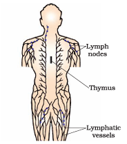
In the provided diagram of the lymphatic system, the small solid structures distributed along the vessels function primarily to:
QUESTION 4 OF 20
While the structures highlighted in Figure 7.5 trap antigens in the lymph, which other secondary lymphoid organ acts similarly but serves as a filter of the blood by trapping blood-borne micro-organisms?
QUESTION 5 OF 20
Which of the following statements is NOT accurate regarding the immune response mediated during an allergy?
QUESTION 6 OF 20
Match the following biochemicals/drugs with their specific roles in allergic reactions:
| Column 1 | Column 2 |
|---|---|
| 1. Histamine | i. Drugs that quickly reduce the symptoms of allergy |
| 2. IgE | ii. Chemicals released from mast cells causing symptoms |
| 3. Anti-histamine | iii. Antibodies produced against specific allergens |
QUESTION 7 OF 20
In higher vertebrates, the development of auto-immune diseases represents a fundamental failure of which advanced immunological ability?
QUESTION 8 OF 20
Consider the following statements regarding rheumatoid arthritis:
I. It is a condition where the body mistakenly attacks its own self-cells.
II. It is a classic example of an auto-immune disease affecting many people in society.
III. It is primarily caused by an infection of the synovial joints by a retrovirus.
Which of the above statements are correct?
QUESTION 9 OF 20
Why is the bone marrow classified as a primary lymphoid organ alongside the thymus?
QUESTION 10 OF 20
Which of the following statements does NOT logically describe the thymus in the context of human aging and immunity?
QUESTION 11 OF 20
Arrange the following biological structures associated with HIV from the innermost genetic core to the outer structure based on the characteristics of a retrovirus:
1. Viral envelope
2. RNA genome
3. Protein coat
QUESTION 12 OF 20
Which of the following groups are identified as being at high risk of getting HIV infection?
I. Individuals who have multiple sexual partners
II. Drug addicts who take drugs intravenously
III. Individuals who require repeated blood transfusions
IV. People who share utensils with an infected person
QUESTION 13 OF 20
If the enzyme reverse transcriptase was completely inhibited in a cell exposed to HIV, what would be the immediate consequence on the viral replication cycle?
QUESTION 14 OF 20
Regarding the role of macrophages in HIV infection, which of the following statements is NOT true?
QUESTION 15 OF 20
During the progression of HIV infection, the patient starts suffering from opportunistic infections (like Mycobacterium or Toxoplasma). What is the primary immunological reason for this vulnerability?
QUESTION 16 OF 20
Arrange the clinical steps of a suspected AIDS case from initial infection to diagnosis:
1. Time-lag with no immediate symptoms.
2. HIV enters helper T-lymphocytes leading to their progressive decrease.
3. Patient suffers bouts of fever, diarrhoea, and weight loss.
4. Diagnosis confirmed using Enzyme Linked Immuno-Sorbent Assay (ELISA).
QUESTION 17 OF 20
Match the carcinogen type to its appropriate example as given in the text:
| Column 1 | Column 2 |
|---|---|
| 1. Physical ionizing radiation | i. Oncogenic viruses |
| 2. Physical non-ionizing radiation | ii. UV rays |
| 3. Chemical carcinogen | iii. X-rays |
| 4. Biological agent | iv. Tobacco smoke |
QUESTION 18 OF 20
What is the critical distinction between cellular oncogenes (c-onc) and viral oncogenes?
QUESTION 19 OF 20
Which of the following is NOT correctly matched regarding cancer detection techniques?
QUESTION 20 OF 20
In the treatment of cancer, why are biological response modifiers like α-interferon administered to patients?
Test Complete!
Answer Review
1
Contact inhibition acts as a cellular brake that halts proliferation upon physical contact with adjacent cells. Oncogenic transformation disrupts these intracellular signaling networks, causing cells to ignore density limits. This loss of regulation leads to continuous mitotic cycles, resulting in abnormal cellular masses called tumors.
In healthy biological tissues, cell proliferation and differentiation are kept in a precise homeostatic balance through a process called contact inhibition. When normal cells divide and physically crowd together, surface-bound cell adhesion molecules trigger intracellular signaling pathways that arrest the cell cycle in the G₁ phase, preventing overcrowding. As detailed in the passage, cancer cells lose this regulatory mechanism. Without contact inhibition, the cells ignore density-dependent signals and divide continuously. They pile up into disorganized, multilayered structures rather than a controlled monolayer. This unchecked mitotic activity forms an abnormal tissue mass known as a tumor or neoplasm, directly validating Option B.
- Option A → Reason: Cancer cells do not cause the immediate death of surrounding normal tissue cells upon contact; instead, they slowly outcompete healthy cells for local nutrients and physical space.
- Option C → Reason: The breakdown of contact inhibition is a failure of internal growth regulation, not a mechanism that activates the immune system to destroy foreign cells.
- Option D → Reason: Converting an RNA virus into a DNA genome is a genetic process carried out by the viral enzyme reverse transcriptase; it is entirely unrelated to physical contact inhibition.
Used: Contextual/Tonal Matching
Application: Match the core phrase in the question directly to the cause-and-effect statement provided in the reading passage. The text states: "As a result of this, cancerous cells just continue to divide giving rise to masses of cells called tumors."
Final Logic: The literal wording in the passage links the loss of contact inhibition directly to tumor formation, pointing to Option B.
Contact Lost = Control Lost →Tumor.
2
Benign tumors stay confined inside a localized fibrous capsule without spreading. Malignant cells degrade extracellular matrix barriers to invade nearby blood and lymph vessels. Migrating tumor cells travel through the blood to form new secondary tumors in distant organs.
Tumors are categorized as either benign or malignant based on their structural growth patterns and ability to spread. Benign tumors stay restricted to their site of origin and do not invade nearby tissues. In contrast, malignant tumors are aggressive masses of neocellular tissue that grow rapidly and invade surrounding structures. Metastasis is a defining, advanced feature of malignant cancers. Cells continuously detach, or slough off, from the primary tumor mass and penetrate nearby blood vessels or lymphatic channels. These circulating tumor cells travel through the vascular network until they lodge in distant capillary beds, such as in the lungs, liver, or brain. They then migrate out of the vessels and begin dividing anew, forming destructive secondary tumors throughout the body. This systemic spread makes treatment difficult, which is why it is considered the most dangerous property of cancer.
- Option A → Reason: Metastasis involves the loss of normal growth controls; it does not cause cancer cells to rapidly regain contact inhibition.
- Option C → Reason: Remaining confined to the original location is a defining characteristic of benign tumors, not spreading malignant ones.
- Option D → Reason: Metastasis is a physical cellular migration process; it does not alter genetics or transform viral oncogenes into cellular oncogenes.
Used: Substitution
Application: Swap the technical term "metastasis" with its literal definition as detailed in the final sentence of the passage.
Final Logic: The text states that cells slough off, travel through the blood, and start new tumors at distant sites, which matches Option B.
Metastasis = Movement (Malignant cells Migrating to distant sites).
3 In the provided diagram of the lymphatic system, the small solid structures distributed along the vessels function primarily to:
Lymph nodes are secondary lymphoid organs placed along lymphatic pathways. They act as physical filters that screen interstitial lymph fluid before it returns to the blood. Trapped pathogens activate local lymphocytes to trigger an adaptive immune response.
The small, solid oval structures located along the lymphatic vessels are lymph nodes, which serve as secondary lymphoid organs. Interstitial fluid that bathes body tissues drains into lymphatic capillaries as lymph, often carrying foreign antigens, cell debris, or pathogens from local infections. As this fluid flows through the lymph nodes, their internal mesh-like structural tissue acts as a physical filter to trap these microorganisms. The nodes are packed with mature B-lymphocytes, T-lymphocytes, and antigen-presenting macrophages. When these immune cells encounter the trapped antigens, they undergo rapid activation and proliferation. This process mounts an adaptive immune response to neutralize the infection before the filtered lymph fluid is returned to the cardiovascular circulatory system.
- Option A → Reason: Erythrocytes (red blood cells) are produced through erythropoiesis inside the red bone marrow, not within the lymphatic vessels or nodes.
- Option B → Reason: While activated plasma cells inside lymph nodes produce immunoglobulins, they do not directly secrete antibodies into the bloodstream; instead, antibodies enter the blood downstream via the thoracic duct.
- Option D → Reason: Immature T-cell progenitors are produced in the bone marrow and migrate to the thymus for maturation, not to the lymph nodes.
Used: Elimination
Application: Identify the structures as lymph nodes and rule out options that describe primary lymphoid functions (production or maturation) or circulatory system functions.
Final Logic: This rules out red blood cell production (A) and T-cell maturation (D), leaving Option B as the correct statement for secondary lymphoid tissue function.
Lymph Nodes = Net (Trapping pathogens in the lymph fluid).
4 While the structures highlighted in Figure 7.5 trap antigens in the lymph, which other secondary lymphoid organ acts similarly but serves as a filter of the blood by trapping blood-borne micro-organisms?
The spleen is a large, bean-shaped secondary lymphoid organ located in the upper abdomen. It filters circulating blood rather than interstitial lymph fluid. Phagocytic cells in the spleen destroy blood-borne pathogens and worn-out erythrocytes.
While lymph nodes filter interstitial tissue fluid, the spleen serves as the primary filter for the cardiovascular circulatory system. The spleen is a large, bean-shaped secondary lymphoid organ located in the upper left quadrant of the abdominal cavity. It is divided structurally into red pulp and white pulp. The white pulp contains organized clusters of B and T lymphocytes that monitor the blood for systemic infections. The red pulp consists of vascular cords packed with macrophages and dendritic cells. As blood moves through this tissue, these phagocytic cells trap and destroy blood-borne microorganisms. Additionally, the spleen removes old, damaged red blood cells, acting as a graveyard for erythrocytes and a reservoir for platelets. [ SECONDARY LYMPHOID FILTERS ] / \ [ Lymph Nodes ] [ Spleen ] | | Filters Tissue Fluid Filters Circulating Blood
- Option A → Reason: The thymus is a primary lymphoid organ located in the chest that manages T-lymphocyte maturation; it does not act as a filter for blood-borne antigens.
- Option B → Reason: The bone marrow is a primary lymphoid tissue responsible for generating all blood cells; it does not function as a secondary filter for circulating pathogens.
- Option D → Reason: The thyroid is an endocrine gland located in the neck that secretes metabolic hormones like thyroxine (T4); it has no direct role in immune filtration.
Used: Category Alignment
Application: Group the options by their physiological systems and developmental roles. The thymus and bone marrow are primary lymphoid organs, while the thyroid belongs to the endocrine system.
Final Logic: The spleen is the only secondary lymphoid organ listed that filters blood, matching the requirements of the question.
Spleen = Screen for Blood.
5 Which of the following statements is NOT accurate regarding the immune response mediated during an allergy?
Allergies are hypersensitivity reactions to typically harmless environmental antigens. Plasma cells produce allergen-specific IgE antibodies are produced during initial sensitization. Antibodies act as molecular adaptors that trigger cellular responses; they cannot engulf or phagocytose particles.
Statement D is factually incorrect because antibodies are soluble proteins, not cells, and therefore cannot perform phagocytosis. Phagocytosis is an active process in which specialized immune cells, such as macrophages and neutrophils, engulf and digest large foreign particles. During an allergic reaction, IgE antibodies function as biochemical adaptors. The constant (Fc) region of the IgE molecule binds to high-affinity FcεRI receptors on the surface of mast cells and basophils. When an individual is re-exposed to the allergen, the allergen binds to and cross-links these surface-bound IgE antibodies. This cross-linking signals the mast cell to degranulate, releasing inflammatory mediators such as histamine into the surrounding tissue. Because antibodies bind to antigens rather than engulfing them, statement D is incorrect and is therefore the correct answer for this negative question.
- Option A → Reason: This is a correct statement. An allergy is defined as an inappropriate, hypersensitive, and exaggerated immune response to otherwise harmless environmental substances.
- Option B → Reason: This is a correct statement. Plasma cells undergo class switching during sensitization to produce Immunoglobulin E (IgE) antibodies.
- Option C → Reason: This is a correct statement. Plant pollen, dust mite feces, mold spores, and animal dander are common environmental allergens.
Used: Elimination
Application: Check the biological definitions in each statement. Phagocytosis is a cellular function that requires a living cell with a cytoskeleton, whereas antibodies are glycoproteins secreted by immune cells.
Final Logic: Since a protein molecule cannot carry out cellular ingestion, statement D contains a clear functional error and must be the incorrect statement.
- t be the intended answer.
Antibodies Bind; Cells Phagocytose.
6 Match the following biochemicals/drugs with their specific roles in allergic reactions:
| Column 1 | Column 2 |
|---|---|
| 1. Histamine | i. Drugs that quickly reduce the symptoms of allergy |
| 2. IgE | ii. Chemicals released from mast cells causing symptoms |
| 3. Anti-histamine | iii. Antibodies produced against specific allergens |
Mast cells release inflammatory chemicals during degranulation to drive symptoms. Specialized immunoglobulins bind to receptor sites to sensitize tissue. Antagonistic medications block target receptors to provide symptomatic relief.
This question matches key molecules and drugs involved in allergic pathways to their functional descriptions: Histamine (1) is an inflammatory chemical mediator stored inside the granules of mast cells and basophils. When released, it binds to local vascular receptors, causing vasodilation, increased capillary permeability, and smooth muscle spasms that lead to allergy symptoms (ii). IgE (2) is the specific antibody class produced by differentiated B-cells during allergic sensitization. It circulates and binds to the surface receptors of mast cells, priming them for subsequent allergen exposure (iii). Anti-histamines (3) are competitive pharmacological antagonists that bind to and block H₁ histamine receptors on target tissues, thereby quickly reducing allergy symptoms such as swelling, sneezing, and itching (i). This yields the correct matching layout: 1-ii, 2-iii, 3-i.
- Option B (1-i, 2-ii, 3-iii) → Reason: This option misidentifies histamine as a therapeutic drug and incorrectly defines IgE as a chemical mediator released from mast cells.
- Option C (1-iii, 2-i, 3-ii) → Reason: This pairing incorrectly suggests that histamine is an antibody, identifies anti-histamines as allergy-inducing chemicals, and mischaracterizes IgE.
- Option D (1-ii, 2-i, 3-iii) → Reason: This combination correctly matches histamine but incorrectly switches the roles of anti-histamine drugs and IgE antibodies.
Used: Option Grouping
Application: Group the items according to their biological functions. The drug anti-histamine (3) acts against histamine (1) to block its effects, matching description i ("Drugs that quickly reduce allergy symptoms"). Therefore, item 3 pairs with i.
Final Logic: Looking at the options, only choices A and D contain the 3-i pairing. Checking item 2 (IgE), it is an antibody (iii), which identifies Option A as the correct combination.
Ig- = Immunoglobulin/Antibody (iii).
7 In higher vertebrates, the development of auto-immune diseases represents a fundamental failure of which advanced immunological ability?
Acquired immunity relies on distinguishing host cells from external pathogens. Immunological tolerance eliminates autoreactive cells during lymphocyte development. A breakdown in this self-tolerance causes the immune system to attack host tissues.
Higher vertebrates have evolved an advanced, memory-based adaptive immune system that relies on distinguishing between "self" markers (the body's own healthy proteins and tissues) and "non-self" markers (foreign pathogens, toxins, and abnormal cells). This self-tolerance is established during lymphocyte development. Immature T and B cells that react too strongly to self-antigens are eliminated or inactivated through clonal deletion and anergy. An autoimmune disease occurs when this self-tolerance mechanism breaks down due to genetic mutations, environmental triggers, or structural similarities between foreign antigens and self-proteins (molecular mimicry). The immune system misidentifies host tissues as foreign threats and mounts an attack against them using autoreactive T-cells and autoantibodies. This represents a failure to differentiate foreign organisms from self-cells, validating Option B.
- Option A → Reason: Patients with autoimmune diseases can still produce primary and secondary immune responses; their immune system is active, but it is misdirected against their own tissues.
- Option C → Reason: Secreting interferons is an innate immune defense used by virus-infected cells to protect neighboring tissue; it is unrelated to adaptive self-tolerance.
- Option D → Reason: Contact inhibition is a structural property of tissue cells that regulates cell density and growth; it is not an immunological mechanism managed by lymphoid cells.
Used: Substitution
Application: Check the baseline definition of autoimmunity in the textbook. The NCERT text states: "The memory-based acquired immunity evolved in higher vertebrates based on the ability to differentiate foreign organisms from self-cells... due to genetic and other unknown reasons, the body attacks self-cells. This results in damage to the body and is called auto-immune disease."
Final Logic: This definition connects the failure to distinguish self from non-self directly to autoimmunity, identifying Option B as correct.
Auto = Self. Autoimmune failure = Attacking Self.
8 Consider the following statements regarding rheumatoid arthritis:
I. It is a condition where the body mistakenly attacks its own self-cells.
II. It is a classic example of an auto-immune disease affecting many people in society.
III. It is primarily caused by an infection of the synovial joints by a retrovirus.
Which of the above statements are correct?
Rheumatoid arthritis is a chronic autoimmune disease that targets joint tissue. Autoantibodies attack the synovial membrane, causing chronic inflammation and damage. The disease is driven by internal immune dysfunction, not an active retroviral infection.
Statement I is correct; rheumatoid arthritis (RA) is a chronic inflammatory disorder in which the immune system mistakenly attacks the body's own tissues. The body produces autoantibodies, such as Rheumatoid Factor (RF), that target its own immunoglobulins, leading to the accumulation of immune complexes in the joints. Statement II is also correct; RA is a classic and common example of an autoimmune disease in humans, causing inflammation, thickening of the synovial membrane, and progressive cartilage damage. Statement III is incorrect; RA is not caused by an active retroviral infection of the joints. Although environmental factors such as smoking or previous viral exposures may contribute to the development of the disease in genetically predisposed individuals, the joint damage itself results from a persistent autoimmune response rather than an active viral infection. Therefore, only Statements I and II are correct.
- Option B (II and III only) → Reason: This option includes the incorrect viral claim in Statement III and excludes Statement I, which correctly describes the underlying autoimmune mechanism.
- Option C (I and III only) → Reason: This choice includes Statement III, which incorrectly identifies a retrovirus as the primary cause of joint damage.
- Option D (I, II, and III) → Reason: This selection incorrectly treats Statement III as true, confusing an autoimmune disease with a direct viral infection.
Used: Elimination
Application: Evaluate the claim made in Statement III. Retroviruses such as HIV cause immunodeficiency disorders, whereas rheumatoid arthritis is classified as an autoimmune disease driven by dysregulation of the immune system.
Final Logic: Since Statement III is factually incorrect, all options containing it (B, C, and D) are eliminated, leaving Option A as the correct answer.
Rheumatoid Arthritis = Rejected Auto-antigens (Statements I and II only).
9 Why is the bone marrow classified as a primary lymphoid organ alongside the thymus?
Primary lymphoid organs are the sites where lymphocytes are generated and mature. Maturation transforms uncommitted progenitor cells into antigen-sensitive cells. Secondary lymphoid organs serve as the sites where these cells interact with foreign antigens.
Lymphoid organs are divided into primary and secondary structures based on their role in lymphocyte development. Bone marrow and the thymus are classified as primary lymphoid organs. These tissues provide specialized microenvironments needed for the early development of immune cells. Inside the bone marrow, hematopoietic stem cells divide and give rise to immature lymphoid progenitors. For B-lymphocytes, the entire process of development, structural gene rearrangement, and maturation occurs within the bone marrow tissue. For T-lymphocytes, immature progenitors travel from the bone marrow to the thymus to complete maturation. In both organs, immature cells mature into functional, antigen-sensitive lymphocytes that express specific surface receptors before entering circulation. This makes Option B the correct explanation. [ PRIMARY LYMPHOID ORGANS ] -----> Maturation & Differentiation (Bone Marrow / Thymus) | v [ SECONDARY LYMPHOID ORGANS ] ----> Antigen Interaction & Proliferation (Spleen / Lymph Nodes)
- Option A → Reason: Providing sites for lymphocytes to interact with antigens is the defining role of secondary lymphoid organs, such as the spleen, lymph nodes, and tonsils.
- Option C → Reason: While red bone marrow contains vascular sinuses that hold red blood cells, storing erythrocytes is a secondary function of the spleen, not the reason it is classified as a primary lymphoid organ.
- Option D → Reason: Histamine is stored and released by tissue mast cells and circulating basophils during inflammatory reactions, not by bone marrow tissue structures.
Used: Category Alignment
Application: Differentiate between primary and secondary lymphoid functions. Primary organs handle lymphocyte production and maturation, while secondary organs manage antigen interaction.
Final Logic: Option A describes a secondary organ function, while Option B correctly describes the maturation role of a primary lymphoid organ.
Primary = Production and Placement of receptors (Maturation). Secondary = Site of Battle.
10 Which of the following statements does NOT logically describe the thymus in the context of human aging and immunity?
The thymus is a primary lymphoid organ located in the chest cavity. It reaches its maximum size relative to body weight around the time of birth. It undergoes age-related involution, shrinking after puberty and reducing new T-cell production.
Statement C is factually incorrect because the thymus does not keep growing past puberty; instead, it undergoes a process called age-related involution. The thymus is a bilobed primary lymphoid organ located in the chest, sitting just behind the sternum (breastbone) and in front of the heart. It provides the specialized microenvironment of epithelial cells and cytokines needed for immature T-lymphocyte progenitors to develop into mature, functional T-cells. The thymus is largest relative to body weight at birth and reaches its absolute maximum physical weight during puberty. After puberty, sex hormone production triggers age-related involution, causing the functional cortical and medullary tissues to gradually shrink and be replaced by fatty adipose tissue. This leads to a decline in the output of new naive T-lymphocytes in older adults. Because statement C describes a pattern of continuous growth that does not occur, it is the correct choice for this "NOT" question.
- Option A → Reason: This is a correct anatomical description. The thymus is situated in the superior mediastinum, beneath the sternum and near the heart.
- Option B → Reason: This is a correct functional description. The thymus provides the microenvironment needed for the selection and maturation of T-lymphocytes.
- Option D → Reason: This is a correct developmental description. The thymus shrinks significantly after puberty, becoming a small tissue remnant in older individuals.
Used: Extreme Word Filter
Application: Look for contradictions between options that describe the same trend. Option C states that the thymus continually increases in size, while Option D states that it reduces to a very small size after birth and puberty.
Final Logic: Since these two growth patterns contradict each other, one must be wrong. Textbook facts confirm that the thymus shrinks over time, making Option C the false statement.
Thymus Time: T-cells mature here, but it shrinks over Time.
11 Arrange the following biological structures associated with HIV from the innermost genetic core to the outer structure based on the characteristics of a retrovirus:
1. Viral envelope
2. RNA genome
3. Protein coat
HIV is an enveloped retrovirus containing a core genetic payload. The absolute center contains two identical single-stranded RNA filaments. This genetic core is enclosed by a protective protein capsid, which is surrounded by an outer lipid membrane.
- Structurally, a retrovirus particle like HIV is built in concentric layers. Looking from the inside out: Innermost Core (2): Consists of the viral RNA genome along with essential enzymes like reverse transcriptase. Middle Layer (3): The genetic material is housed directly within an icosahedral protein coat (capsid). Outermost Layer (1): Enclosing the protein coat is the viral envelope, a lipid bilayer derived from the host cell's plasma membrane embedded with viral glycoproteins. Arranging these architectural components from innermost to outermost gives the sequence 2 →3 →1, matching option A.
- Option B → Falsely places the viral envelope (1) at the innermost center and the RNA genome (2) on the outside.
- Option C → Places the protein coat (3) at the absolute center, which is structurally incorrect since it encapsulates the RNA.
- Option D → Incorrectly sandwiches the outer viral lipid envelope (1) between the internal genome and the capsid coat.
Used: Elimination
Application: Identify the absolute boundaries. The genetic material (RNA genome, 2) must be at the very center, and the envelope membrane (1) must form the exterior boundary. This means the sequence must start with 2 and end with 1.
Final Logic: Option A is the only sequence that positions the genome at the absolute core and the envelope on the outside.
Inside-Out HIV: RNA →Protein Capsid →Envelope (Read Protein Everywhere).
12 Which of the following groups are identified as being at high risk of getting HIV infection?
I. Individuals who have multiple sexual partners
II. Drug addicts who take drugs intravenously
III. Individuals who require repeated blood transfusions
IV. People who share utensils with an infected person
HIV transmission requires the direct exchange of infected body fluids. High-risk behaviors include unprotected sex with multiple partners and sharing needles. Medical vulnerabilities include relying on frequent blood transfusions from un-screened donors.
- HIV is not a highly contagious airborne or casual contact virus. It spreads through specific fluids like blood, semen, and vaginal secretions. Statements I, II, and III represent well-established transmission routes: multiple sexual partners increase exposure risk; shared needles expose drug users to infected blood; and frequent transfusions increase the probability of encountering contaminated blood units. Statement IV is false: HIV cannot survive outside the body on dry utensils, nor is it transmitted through saliva via sharing plates or cups. Casual, everyday social interactions pose zero risk. Therefore, only statements I, II, and III are valid high-risk factors.
- Option A → Includes statement IV, falsely implying that casual contact via sharing household utensils can transmit HIV.
- Option B → Includes statement IV and leaves out the highly critical sexual transmission vector covered in statement I.
- Option D → Incorrectly includes statement IV while omitting the high-risk intravenous drug usage category described in statement II.
Used: Extreme Word Filter / Fact Check
Application: Isolate statement IV ("share utensils"). This is a classic medical misconception. NCERT explicitly states that HIV does not spread by casual contact. Eliminating any option containing IV automatically reveals the correct choice.
Final Logic: Removing IV eliminates options A, B, and D, leaving option C as the correct answer.
Fluids, Not Food: HIV travels through biological fluids (Fluids), not through sharing food/forks (Food).
13 If the enzyme reverse transcriptase was completely inhibited in a cell exposed to HIV, what would be the immediate consequence on the viral replication cycle?
Retroviruses like HIV store their genetic information as single-stranded RNA. Inside the host cell, reverse transcriptase builds a complementary DNA strand from this RNA template. Inhibiting this enzyme stops the viral genetic material from converting into a DNA form.
- The defining feature of a retrovirus is its ability to reverse the standard flow of genetic information (which normally moves from DNA to RNA). Once the HIV core enters a host cell, it uses its viral enzyme, reverse transcriptase, to rewrite its single-stranded viral RNA into double-stranded viral DNA. If a pharmacological agent or mutation completely blocks this enzyme, this conversion fails entirely. The virus cannot generate viral DNA, preventing it from integrating into the host's genome or producing new viral copies.
- Option A → Viral binding to a macrophage depends on the outer glycoprotein gp120 docking with host CD4 receptors; this happens before reverse transcriptase is released.
- Option C → Without the enzyme, viral DNA cannot be synthesized in the first place, making any over-replication or lysis impossible.
- Option D → While the ultimate lack of new virus production protects helper T-cells, this is a downstream result. The immediate cellular consequence is the failure to make viral DNA.
Used: Contextual/Tonal Matching
Application: Match the function of the enzyme directly to its biological definition. "Reverse transcriptase" means transcribing backward: RNA →DNA. Look for the choice that describes a breakdown in this exact biochemical conversion step.
Final Logic: Option B directly describes the failure of RNA transforming into viral DNA, which matches the definition of reverse transcriptase inhibition.
Reverse Transcriptase = RNA to DNA: Block the enzyme, block the biochemical bridge to DNA.
14 Regarding the role of macrophages in HIV infection, which of the following statements is NOT true?
Macrophages are among the first immune cells targeted by HIV upon entering the body. Unlike helper T-cells, macrophages are not rapidly lysed or destroyed by viral replication. They survive long-term, continuously shedding new viral particles into the system.
- When HIV enters the human body, it targets cells expressing the CD4 receptor, primarily macrophages and helper T-lymphocytes (TH cells). Inside macrophages, the virus converts its RNA into DNA, integrates into the host genome, and directs the cell to assemble new virions. Crucially, macrophages can tolerate viral replication without dying immediately. They survive for extended periods, functioning as persistent reservoirs that continuously release new viruses, earning them the nickname "HIV factories." Statement C claims that macrophages are destroyed immediately, which is factually incorrect and therefore the correct choice for this "NOT true" question.
- Option A → Reason: This statement is true. Macrophages are among the first host cells infected during the early stages of HIV infection.
- Option B → Reason: This statement is true. The conversion of viral RNA into viral DNA by reverse transcriptase occurs within the cytoplasm of the infected macrophage.
- Option D → Reason: This statement is true. Because macrophages survive infection and continuously produce progeny virions, they are often described as "HIV factories" in biology textbooks and reference materials.
Used: Odd One Out / Concept Contradiction
Application: Analyze options C and D. They directly contradict each other. A cell cannot be "destroyed immediately" (C) if it simultaneously "continues to produce virus as a factory" (D). Recognizing from NCERT that macrophages act as long-term factories helps you identify statement C as the false one.
Final Logic: Since macrophages survive the infection to continuously shed virus, statement C is false and satisfies the question's condition.
T-Cells = Terminated (Numbers drop drastically).
15 During the progression of HIV infection, the patient starts suffering from opportunistic infections (like Mycobacterium or Toxoplasma). What is the primary immunological reason for this vulnerability?
Helper T-lymphocytes (TH cells) coordinate the immune system's response to pathogens. HIV actively infects, replicates within, and destroys these helper T-cells. When T-cell counts fall below critical levels, the immune system can no longer effectively defend the body against common pathogens.
Helper T-lymphocytes (CD4⁺ T-cells) act as the central coordinators of adaptive immunity, releasing cytokines that activate both B-cells (for antibody production) and cytotoxic T-cells (for cell-mediated defense). As HIV infection progresses, the virus systematically infects and destroys these helper T-cells. Over time, their numbers decline drastically. This loss leaves the immune system severely compromised, resulting in a profound state of immunodeficiency. Common environmental pathogens—such as Mycobacterium, Toxoplasma, fungi, and viruses—that are normally controlled by a healthy immune system can then cause serious and potentially life-threatening opportunistic infections.
- Option A → Overproduction of IgE antibodies is a characteristic feature of allergic responses, not a mechanism of HIV-induced immunodeficiency.
- Option C → HIV is a retrovirus that disrupts immune signaling by destroying specific cells; it does not produce or secrete physical chemical toxins to paralyze bone marrow tissue.
- Option D → Loss of contact inhibition is a hallmark of cancerous cells turning into tumors, not a mechanism associated with HIV viral replication or immunodeficiency.
Used: Contextual/Tonal Matching
Application: Match the acronym AIDS (Acquired Immuno-deficiency Syndrome) with the options. Look for the choice that explains a profound loss of immune defense. A massive drop in helper T-cells (Option B) explains this systemic immune collapse.
Final Logic: The defining clinical cause of opportunistic infections in AIDS patients is the destruction and drop in helper T-lymphocytes.
Helper T-cells = Help is gone: When HIV destroys the Helper cells, the body has no Help left to fight off basic infections.
16 Arrange the clinical steps of a suspected AIDS case from initial infection to diagnosis:
1. Time-lag with no immediate symptoms.
2. HIV enters helper T-lymphocytes leading to their progressive decrease.
3. Patient suffers bouts of fever, diarrhoea, and weight loss.
4. Diagnosis confirmed using Enzyme Linked Immuno-Sorbent Assay (ELISA).
Initial HIV infection is followed by a long, asymptomatic incubation period (time-lag). Inside the body, the virus silently infects and reduces the helper T-lymphocyte population. As T-cell numbers drop, the patient develops visible clinical symptoms like fever and weight loss. The presence of the disease is finally confirmed through diagnostic tests like an ELISA screen.
- The natural history and clinical presentation of HIV follows a predictable timeline: Step 1: Following exposure, there is a distinct time-lag or incubation period before clear signs appear (ranging from a few months up to 5–10 years). Step 2: During this asymptomatic window, the virus actively replicates inside the lymphoid organs, causing a progressive decline in the host's helper T-lymphocyte count. Step 3: Once the T-cell count falls low enough, the patient develops symptomatic disease, experiencing recurrent fever, chronic diarrhea, and unexplained weight loss. Step 4: The patient seeks medical care, and the disease is formally identified using the standard ELISA screening assay. This logical clinical progression corresponds perfectly to the sequence 1, 2, 3, 4.
- Option A → Falsely places the physiological drop in T-cells (2) before the initial asymptomatic time-lag phase (1), and positions the final confirmation test (4) before symptoms surface (3).
- Option C → Suggests that major clinical wasting symptoms (3) occur before the underlying destruction of helper T-cells (2) has taken place.
- Option D → Proposes an illogical sequence that places symptoms and viral cell entry well before the incubation time-lag phase has elapsed.
Used: Substitution / Chronological Logic
Application: Place the events in order based on a standard medical timeline: Infection →Incubation Window (1) →Cellular Damage (2) →Visible Symptoms (3) →Lab Confirmation (4). This timeline matches option A exactly.
Final Logic: Diagnostic confirmation (4) must be the final step in a clinical workup, meaning the sequence must end with 4, which immediately highlights Option B or C. Ordering the timeline correctly confirms B.
Silent to Symptomatic: Silent lag →Cell drop →Symptoms show →Test to confirm.
17 Match the carcinogen type to its appropriate example as given in the text:
| Column 1 | Column 2 |
|---|---|
| 1. Physical ionizing radiation | i. Oncogenic viruses |
| 2. Physical non-ionizing radiation | ii. UV rays |
| 3. Chemical carcinogen | iii. X-rays |
| 4. Biological agent | iv. Tobacco smoke |
Ionizing radiation like X-rays possesses high energy that breaks chemical bonds and damages DNA. Non-ionizing radiation like ultraviolet (UV) rays has lower energy but can still cause mutational damage. Chemical carcinogens include toxic compounds found in tobacco smoke. Biological oncogenic agents consist of cancer-causing viruses.
- Carcinogens are agents that transform normal cells into cancerous ones. They are categorized by their nature: 1. Physical ionizing radiation matches iii (X-rays): High-energy waves that penetrate deeply and break DNA strands. 2. Physical non-ionizing radiation matches ii (UV rays): Lower-energy waves that damage DNA by causing pyrimidine dimers, often leading to skin cancers. 3. Chemical carcinogen matches iv (Tobacco smoke): Contains harmful chemical compounds that are a leading cause of lung cancer. 4. Biological agent matches i (Oncogenic viruses): Viruses that carry cancer-causing genes (viral oncogenes) into host cells. Matching these up gives 1-iii, 2-ii, 3-iv, 4-i, which corresponds to option C.
- Option A → Mismatches physical non-ionizing radiation (2) with tobacco smoke (iv), which is a chemical agent.
- Option B → Incorrectly switches the physics terms, calling UV rays ionizing (1-ii) and X-rays non-ionizing (2-iii).
- Option D → Incorrectly lists ionizing radiation (1) as tobacco smoke (iv) and biological agents (4) as X-rays (iii).
Used: Option Grouping / Physics Anchors
Application: Anchor your matching on basic physics: X-rays are high-energy ionizing radiation (1 matches with iii), while UV rays are non-ionizing radiation (2 matches with ii). Look for an option starting with 1-iii and 2-ii.
Final Logic: Only option C contains both correct physical radiation matches, making it the correct answer.
Non-ionizing = Natural (UV rays from the sun).
18 What is the critical distinction between cellular oncogenes (c-onc) and viral oncogenes?
Normal cells contain inactive genes called cellular oncogenes (c-onc) or proto-oncogenes. When triggered by carcinogens, these c-onc switch on, leading to cancerous transformation. Viral oncogenes are actual cancer-causing genes carried and introduced by oncogenic viruses.
- The human genome contains normal genes called proto-oncogenes or cellular oncogenes (c-onc). These genes normally regulate regular cell growth and division. However, if they are mutated or activated abnormally by environmental carcinogens, they can drive the cell toward uncontrolled, cancerous growth. In contrast, viral oncogenes are foreign, cancer-inducing genes carried within the genetic payload of oncogenic viruses. When these viruses infect a host cell, they insert these viral oncogenes to directly force neoplastic transformation. Option B outlines this genetic distinction.
- Option A → Falsely claims c-onc are physical radiations and viral oncogenes are chemicals; both are types of genes, not physical or chemical agents.
- Option C → Proto-oncogenes do not inhibit cell division; they are positive regulators of normal growth that cause over-activation when mutated.
- Option D → Both gene types are involved in driving the development of malignant tumors; they are not separated by benign versus malignant tumor classifications.
Used: Contextual/Tonal Matching
Application: Look at the names of the genes. "Cellular oncogene" implies it belongs to the host's own cells. "Viral oncogene" implies it originates from a virus. Option B is the only choice that matches this naming logic.
Final Logic: Option B correctly defines the cellular origins of c-onc and the viral source of viral oncogenes based on NCERT guidelines.
Viral oncogene = Vector/Virus (brought in from the outside).
19 Which of the following is NOT correctly matched regarding cancer detection techniques?
Biopsies and blood tests rely on cellular examination under a microscope. Computed Tomography (CT) scans use X-ray radiation to create 3D internal views. Magnetic Resonance Imaging (MRI) uses non-ionizing radio waves and strong magnetic fields, avoiding dangerous radiation.
- Magnetic Resonance Imaging (MRI) is a diagnostic technique used to safely image internal tissues. It relies on a strong magnetic field and non-ionizing radiofrequency pulses to change the alignment of hydrogen nuclei in the body. It does not use ionizing radiation (like X-rays or gamma rays), which makes it a safer choice for repeated scans. Because Option C states that MRI uses ionizing radiation, it is factually incorrect and the correct answer for this "NOT correctly matched" question.
- Option A → This is a correct match; a biopsy involves taking a small tissue sample, slicing it thin, staining it, and examining it under a microscope (histopathology).
- Option B → This is a correct match; CT scans use rotational X-ray beams and computer processing to create cross-sectional 3D images of internal anatomy.
- Option D → This is a correct match; leukemia is a blood cancer characterized by an abnormal spike in white blood cell counts, which is detected via complete blood counts and bone marrow aspirates.
Used: Extreme Word Filter / Fact Check
Application: Look closely at the words used in Option C: "MRI: Uses ionizing radiations". The word "Magnetic" in MRI tells you it relies on magnetic fields and radio waves, which are non-ionizing. This makes option C incorrect, meaning it is the mismatched answer we are looking for.
Final Logic: Since MRIs are safe because they do not use ionizing radiation, statement C is false and the correct choice.
CT = Cross-section with Corrosive/ionizing radiation (X-rays).
20 In the treatment of cancer, why are biological response modifiers like α-interferon administered to patients?
Tumor cells develop mechanisms to hide from host immune surveillance. Biological response modifiers are immunotherapy tools that help the body recognize these hidden cells. Administering α-interferon alerts and activates the immune system to locate and destroy the tumor cells.
- Cancer cells are mutated versions of the body's own cells, allowing them to dodge detection by the immune system. To counter this camouflage, cancer therapies often include immunotherapy alongside surgery or chemotherapy. α-interferon is a natural biological response modifier. When given to a patient, it stimulates natural killer (NK) cells, macrophages, and T-lymphocytes. This activation helps the immune system spot the hidden cancer cells and mount an effective defense to destroy the tumor.
- Option A → Interferons are immune signaling proteins that boost white blood cells; they do not physically compress or chemically dissolve tumor masses on their own.
- Option C → The loss of contact inhibition is caused by deep genetic damage inside the cancer cells; interferons cannot repair these mutations or restore normal cellular growth breaks.
- Option D → Chemotherapy drugs target and disrupt the cell division machinery during mitosis. Interferon works through immunotherapy, not direct cytotoxicity.
Used: Contextual/Tonal Matching
Application: Connect the term "Biological Response Modifier" with the options. This term means modifying the body's natural biological response—its immune defense. Option B is the only choice that focuses on activating the immune system to fight the tumor.
Final Logic: α-interferon is specifically classified in NCERT as an immunotherapeutic biological response modifier that helps the immune system target tumors.
Interferon = Interfere with evasion: Interferon interferes with the tumor's ability to hide, pointing them out to the immune system.