
CUET UG Biology Booster Test 3-Immune Disorders AIDS and Cancer
📌 Answers are locked once submitted — results and explanations appear at the end.
QUESTION 1 OF 20
QUESTION 2 OF 20
QUESTION 3 OF 20
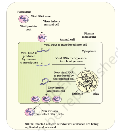
Based on the viral replication diagram, if a highly specific pharmacological drug successfully destroys the newly synthesized viral DNA immediately after its formation in the cytoplasm, which subsequent step in the cycle will be directly prevented?
QUESTION 4 OF 20
The text notes that macrophages act as "HIV factories" while helper T-lymphocytes experience a progressive decrease in number. Analytically, what does the "factory" role imply about the viral replication cycle in macrophages compared to T-cells?
QUESTION 5 OF 20
Match the specific immunological component with its corresponding clinical manifestation or cellular role:
| Column 1 | Column 2 |
|---|---|
| 1. IgE antibodies | i. Rejection of organ transplants |
| 2. Histamine | ii. Rheumatoid arthritis |
| 3. T-lymphocytes (cell-mediated) | iii. Triggers allergic symptoms from mast cells |
| 4. Auto-antibodies | iv. Exaggerated response to pollen or dust |
QUESTION 6 OF 20
Arrange the physiological events of an allergic reaction in their correct chronological order:
1. Release of histamine and serotonin from mast cells.
2. Exposure of the host to an environmental allergen (e.g., pollen).
3. Production of highly specific IgE antibodies.
4. Appearance of clinical symptoms like sneezing and wheezing.
QUESTION 7 OF 20
Which of the following clinical scenarios does NOT represent an auto-immune response based on the fundamental definition provided?
QUESTION 8 OF 20
Rheumatoid arthritis is classified as an auto-immune disease. What fundamental property of memory-based acquired immunity is pathologically bypassed in patients suffering from this condition?
QUESTION 9 OF 20
Consider the following analytical statements regarding the spleen:
I. It acts primarily as a primary lymphoid organ for the maturation of B-cells.
II. It traps blood-borne micro-organisms, acting as a crucial filter for the blood.
III. It serves as a large reservoir of erythrocytes.
Which of the statements given above are correct?
QUESTION 10 OF 20
If a pathogen directly invades through the mucosal epithelium of the respiratory tract, which specific component of the lymphoid tissue is anatomically positioned to encounter it first before it reaches systemic circulation?
QUESTION 11 OF 20
Which of the following analytical statements about the HIV pathogen is INCORRECT?
QUESTION 12 OF 20
Why is HIV/AIDS transmission restricted to specific high-risk behaviors and not spread by mere physical touch?
QUESTION 13 OF 20
An AIDS patient rarely dies directly from the cytopathic effects of the HIV virus itself. Based on the pathogenesis of the disease, what is the most accurate explanation for the high mortality rate?
QUESTION 14 OF 20
Arrange the following clinical steps from the earliest diagnostic intervention to late-stage disease management:
1. Detection of a progressive, severe decrease in Helper T-cells.
2. Initial diagnosis of suspected infection using the Enzyme Linked Immuno-Sorbent Assay (ELISA).
3. Prolongation of the patient's life using anti-retroviral drugs.
4. Onset of severe opportunistic infections like Toxoplasma.
QUESTION 15 OF 20
Which of the following is NOT a logical downstream consequence of the loss of contact inhibition in human cells?
QUESTION 16 OF 20
In the context of malignant tumors, why do neoplastic cells effectively starve normal surrounding cells of vital nutrients?
QUESTION 17 OF 20
Consider the following statements regarding carcinogens and oncogenesis:
I. Non-ionizing radiations like UV rays cause DNA damage leading to neoplastic transformation.
II. Chemical carcinogens present in tobacco smoke have been firmly identified as a major cause of lung cancer.
III. Oncogenic viruses initiate cancer solely by activating the host's existing cellular oncogenes (c-onc).
Which of the statements given above are analytically correct?
QUESTION 18 OF 20
Match the genetic elements with their role in oncogenesis:
| Column 1 | Column 2 |
|---|---|
| 1. Cellular oncogenes (c-onc) | i. Agents (physical/chemical/biological) that induce DNA damage or trigger transformation |
| 2. Viral oncogenes | ii. Genes carried into the host by cancer-causing viruses |
| 3. Carcinogens | iii. Genes identified in normal cells that cause transformation when aberrantly activated |
QUESTION 19 OF 20
A patient is suspected of having leukemia. Why would a blood and bone marrow test be prioritized over a CT scan for the initial histopathological diagnosis?
QUESTION 20 OF 20
Which of the following is NOT an accurate rationale for utilizing a combination of surgery, radiotherapy, and biological response modifiers in modern cancer treatment?
Test Complete!
Answer Review
1
The bone marrow is the primary site of hematopoiesis, where all types of blood cells originate. Both B-lymphocytes and T-lymphocytes are produced initially within the bone marrow from hematopoietic stem cells. A non-functional bone marrow creates a baseline manufacturing failure, completely cutting off the production of immature lymphocytes.
- The bone marrow is designated as the primary primary lymphoid organ because it serves as the foundational manufacturing facility for the entire circulatory and immune systems. Every line of blood cellular element—including erythrocytes, leukocytes, platelets, and the common lymphoid progenitors that give rise to B and T-lymphocytes—is produced inside the red bone marrow. If a genetic defect completely neutralizes bone marrow function from birth, the upstream production of these cells ceases immediately. Without immature lymphocytes entering the pipeline, downstream immunological milestones like thymic maturation, peripheral migration, or antigen activation cannot occur. Thus, option B highlights the absolute baseline failure that happens first.
- Option A → Spleen filtration is a downstream secondary lymphoid event. While it would eventually lack working lymphocytes to mount active defenses, the mechanical filtering mechanism itself is not the first foundational element broken at the cellular birth site.
- Option C → Mast cells release histamine during allergies, but this mechanism is a specialized peripheral allergic response downstream from general immune cell line generation.
- Option D → T-lymphocytes must first be produced by the bone marrow and matured by the thymus before they can gather inside lymph nodes for activation; this step is downstream from basic cell production.
Used: Elimination
Application: Evaluate the anatomical hierarchy of the lymphoid system. Bone marrow is the ultimate source. If the ultimate source/factory is missing, the very first consequence must be a lack of basic materials (cells), rather than secondary tissue operations or activation phases.
Final Logic: Bone marrow is defined by NCERT as the main organ where all blood cells are produced, making its absolute manufacturing failure the first immediate consequence.
Marrow = Maker: The bone Marrow is the cell Maker; no marrow means no immune cell components are created.
2
The thymus provides the specialized micro-environment required for the differentiation and maturation of T-lymphocytes. The thymus reaches its maximum relative size during early life and undergoes progressive atrophy (shrinks) after puberty. Due to this shrinking, elderly individuals experience a decline in their capacity to mature new, naive T-lymphocyte populations.
- The thymus follows a distinct timeline called age-associated involution. It is highly active throughout infancy and early childhood, providing the signaling environment needed to train immature lymphocytes into functional, antigen-sensitive T-cells. Post-puberty, the functional tissue steadily undergoes fatty replacement and shrinks significantly. By old age, the output of newly matured, naive T-cells drops sharply. This loss reduces cell-mediated immune adaptability, leaving elderly individuals more vulnerable to new infections and reducing their vaccine responses compared to young children.
- Option A → The thymus steadily shrinks (atrophies) instead of expanding as a person ages, which limits rather than boosts naive T-lymphocyte production.
- Option C → Age-related involution leads to a loss of tissue function rather than overactivity or growth of the thymic cortex.
- Option D → While the bone marrow continues to produce immature T-cell precursors throughout a person's life, it cannot replace the specialized epithelial environment of the thymus needed to mature those cells.
Used: Contextual/Tonal Matching
Application: The passage states that the thymus "keeps reducing in size with age" and drops to a "very small size" by puberty. The answer choice must directly connect this physical shrinking with a drop in the thymus's primary function: maturing T-lymphocytes.
Final Logic: A tiny, atrophied organ cannot perform large-scale maturation work, which directly supports the conclusion in Option B.
Tiny Thymus = Tired T-cells: A shrinking Thymus yields fewer new T-cells over Time.
3 Based on the viral replication diagram, if a highly specific pharmacological drug successfully destroys the newly synthesized viral DNA immediately after its formation in the cytoplasm, which subsequent step in the cycle will be directly prevented?
Reverse transcriptase synthesizes viral DNA from the viral RNA template inside the host cell's cytoplasm. Once formed, this viral DNA travels into the nucleus to integrate into the host's genome. Destroying this viral DNA right after it forms directly stops it from integrating into the host DNA.
- The replication cycle of a retrovirus follows a specific sequence of steps: binding and entry →uncoating →reverse transcription of viral RNA into viral DNA →integration into host DNA →transcription and translation of viral parts →assembly and budding. If a drug targets and breaks down the newly generated viral DNA right after reverse transcription finishes in the cytoplasm, the virus cannot take its next step: transporting that DNA into the nucleus and using the integrase enzyme to patch it into the host's chromosome. This breaks the lifecycle right before genomic integration occurs.
- Option A → The uncoating of the capsid occurs earlier in the cycle, right after entry and well before reverse transcriptase begins making viral DNA.
- Option C → The initial attachment and membrane fusion steps happen on the outer cell surface, long before any viral DNA is assembled inside the cytoplasm.
- Option D → The release of the core genomic single-stranded RNA into the cytoplasm is a prerequisite step that must happen before reverse transcription can begin.
Used: Sequence/Chronological Logic
Application: Map out the sequential steps of the retroviral lifecycle. The step that directly follows "formation of viral DNA" is "integration of viral DNA into the host genome." This means blocking the DNA stops the integration step that comes next.
Final Logic: Since integration relies entirely on the presence of viral DNA, destroying that DNA directly blocks Option B.
No Copy, No Paste: If you destroy the new viral DNA code (No Copy), the virus cannot integrate it into the host cell's genome (No Paste).
4 The text notes that macrophages act as "HIV factories" while helper T-lymphocytes experience a progressive decrease in number. Analytically, what does the "factory" role imply about the viral replication cycle in macrophages compared to T-cells?
In helper T-cells, rapid viral replication leads to cell lysis and a drop in cell counts. In contrast, infected macrophages act as long-term, stable hosts for the virus. Macrophages continuously produce and bud off new virions without dying, working like a factory.
- The difference in how HIV affects macrophages versus helper T-lymphocytes (Tₕ cells) determines how the disease progresses. When HIV replicates inside helper T-cells, it destroys them, causing a severe drop in cell counts over time. However, when the virus infects a macrophage, it uses the cell's machinery to assemble and bud off new viral particles continuously without causing immediate cell death or lysis. This allows the macrophage to survive long-term as a living reservoir that steadily pumps out new viruses into the bloodstream—matching the definition of a "factory."
- Option B → Immediate lysis is what happens to helper T-lymphocytes, which explains why their numbers drop while macrophages survive to keep producing virus.
- Option C → Macrophages readily integrate viral DNA into their own genome, which is exactly how they are transformed into long-term viral production centers.
- Option D → HIV is an immunodeficiency virus that uses host cells to produce new virions; it does not convert its genome into cellular oncogenes to cause cancer.
Used: Contextual/Tonal Matching
Application: Think about what a "factory" does: it remains standing and operational while continuously manufacturing goods over time. Option A matches this definition by explaining that the cell survives to keep producing new viruses.
Final Logic: The long-term survival and steady production of viruses by macrophages is why NCERT describes them as "HIV factories," making A the correct answer.
Factories Stay Open: Macrophages stay open for business (Maintained factories), while T-cells close down and break (Terminated by lysis).
5 Match the specific immunological component with its corresponding clinical manifestation or cellular role:
| Column 1 | Column 2 |
|---|---|
| 1. IgE antibodies | i. Rejection of organ transplants |
| 2. Histamine | ii. Rheumatoid arthritis |
| 3. T-lymphocytes (cell-mediated) | iii. Triggers allergic symptoms from mast cells |
| 4. Auto-antibodies | iv. Exaggerated response to pollen or dust |
IgE antibodies are the specific class of immunoglobulins produced during an allergic response. Histamine is an inflammatory chemical released by mast cells that drives allergic symptoms. Cell-mediated immunity driven by T-lymphocytes is responsible for graft and organ transplant rejection. Auto-antibodies are produced when self-tolerance fails, leading to diseases like rheumatoid arthritis.
- Let us match each immunological component to its clinical role: 1. IgE antibodies →iv: These are the specific immunoglobulins generated during exaggerated hypersensitivity reactions to environmental antigens like pollen or dust mites. 2. Histamine →iii: This chemical mediator is released from mast cell granules during an allergy, causing localized swelling, irritation, and inflammation. 3. T-lymphocytes →i: Cell-mediated immunity (CMI) is the primary system responsible for identifying foreign tissue grafts and driving organ transplant rejection. 4. Auto-antibodies →ii: These are antibodies that mistakenly target the body's own tissues, leading to chronic auto-immune disorders like rheumatoid arthritis. This complete set matches the 1-iv, 2-iii, 3-i, 4-ii pattern found in option D.
- Option A → Correctly pairs 1 and 2, but incorrectly swaps the roles of T-lymphocytes (3-ii) and auto-antibodies (4-i).
- Option B → Incorrectly matches IgE (1) with mast cell degranulation triggers (iii) and swaps the roles of T-lymphocytes and auto-antibodies.
- Option C → Falsely connects IgE (1) to transplant rejection (i) and incorrectly attributes histamine (2) to rheumatoid arthritis (ii).
Used: Option Grouping / Matching Anchors
Application: Use the most distinct NCERT match as your anchor: T-lymphocytes/cell-mediated immunity is explicitly responsible for graft/transplant rejection (3 matches with i). Check the options where 3 matches with i, which narrows your choices down to Option A or Option D.
Final Logic: Checking the remaining terms shows that auto-antibodies (4) match with rheumatoid arthritis (ii), proving that Option A is the correct configuration.
RA Auto: Rheumatoid Arthritis is driven by Auto-antibodies.
6 Arrange the physiological events of an allergic reaction in their correct chronological order:
1. Release of histamine and serotonin from mast cells.
2. Exposure of the host to an environmental allergen (e.g., pollen).
3. Production of highly specific IgE antibodies.
4. Appearance of clinical symptoms like sneezing and wheezing.
An allergic reaction begins when a person is first exposed to an environmental allergen. This exposure prompts B-cells to produce specific IgE antibodies that bind to tissue mast cells. Re-exposure causes the mast cells to degranulate, releasing inflammatory chemicals like histamine. These free chemicals then trigger visible clinical symptoms such as sneezing and wheezing.
- The development of an allergic reaction follows a step-by-step timeline: Step 1 (Event 2): The host is exposed to an environmental allergen (e.g., breathing in pollen grains). Step 2 (Event 3): The immune system responds to this entry by producing highly specific IgE antibodies, which coat the surface of local mast cells. Step 3 (Event 1): Upon subsequent re-exposure, the allergen cross-links the bound IgE molecules, triggering the release of histamine and serotonin from the mast cells. Step 4 (Event 4): These chemical mediators alter local blood vessels and tissue spaces, causing the appearance of clinical symptoms like sneezing, watering eyes, and wheezing. This chronological sequence corresponds to 2, 3, 1, 4, matching option A.
- Option A → Illogically places the release of mast cell chemicals (1) before the helper B-cells have even synthesized the IgE antibodies (3) needed to trigger degranulation.
- Option B → Falsely suggests that specific IgE antibodies are produced (3) before the host ever encounters the allergen (2).
- Option D → Proposes an inverted sequence where downstream chemical release (1) occurs before the baseline allergen exposure (2) has taken place.
Used: Sequence/Chronological Logic
Application: Determine the absolute beginning and ending steps of the path. An allergy cannot start without initial contact with an allergen (2), and the final result observed is always the visible symptom outbreak (4). The sequence must start with 2 and end with 4.
Final Logic: This narrows the choices to options A and C. Since IgE production (3) must happen before those antibodies can cause mast cells to release chemicals (1), the correct sequence must be 2, 3, 1, 4.
Allergy Axis: Allergen entry →Antibody built →Chemicals clear →Symptoms start.
7 Which of the following clinical scenarios does NOT represent an auto-immune response based on the fundamental definition provided?
Auto-immunity happens when the body's immune system mistakenly attacks its own healthy self-cells. Transplant rejection is a normal, healthy immune response identifying foreign, non-self donor tissue. Since transplant rejection targets external, non-self antigens, it does not fit the definition of an auto-immune disease.
- Auto-immunity is defined as a failure of self-tolerance where the immune system attacks the host's own tissues. Scenarios A, B, and D all fit this definition perfectly. In contrast, when a patient's cell-mediated immune system rejects a transplanted kidney (Scenario C), it is performing its normal job correctly. The cytotoxic T-lymphocytes accurately identify the human leukocyte antigens (HLAs) on the donor kidney as foreign (non-self) and launch an attack to destroy them. Because this response targets a foreign graft rather than the host's own native cells, it is a normal immune reaction, not an auto-immune breakdown.
- Option A → This is a classic description of the root cause of auto-immunity: genetic or environmental failures that disrupt self-tolerance.
- Option B → This describes the physiological impact of auto-immunity, where self-reactive immune cells cause structural damage to the host's own organs.
- Option D → This is a classic example of an auto-immune condition where the body's joints are mistakenly targeted and damaged by its own immune response.
Used: Odd One Out
Application: Group the options based on whether they target internal or external tissues. Options A, B, and D all describe the body attacking its own native cells ("self"). Option C describes an attack on a foreign organ brought in from the outside ("non-self").
Final Logic: Because option C describes a normal defense against foreign tissue rather than an attack on self-cells, it is the odd one out and the correct answer.
Auto means Self: Auto-immune requires an attack on Self-tissue. A transplant from a donor is Non-self.
8 Rheumatoid arthritis is classified as an auto-immune disease. What fundamental property of memory-based acquired immunity is pathologically bypassed in patients suffering from this condition?
Acquired immunity relies on the critical ability to tell the difference between "self" and "non-self." In rheumatoid arthritis, this self-tolerance boundary breaks down entirely. This failure causes the immune system to mistake normal joint tissue for foreign invaders, launching an internal attack.
- Acquired (adaptive) immunity in higher vertebrates has two key features: antigen specificity/memory and self-tolerance. Self-tolerance ensures that as B and T-lymphocytes mature, any cells that react to the body's own proteins are eliminated or turned off. In a patient with rheumatoid arthritis, this protective filter fails. The immune system loses its ability to distinguish foreign pathogens from self-cells, leading auto-reactive antibodies to target and inflame the synovial membranes of the joints.
- Option A → The human immune system produces proteins like antibodies and lysozymes, but it does not synthesize or secrete chemical antibiotic drugs.
- Option C → The skin forms a physical barrier that is part of the innate, non-specific immune system, not an adaptive, memory-based property.
- Option D → Macrophages retain their ability to perform phagocytosis in auto-immune patients; the underlying pathology is a mistake in targeting, not a failure of cell engulfment.
Used: Contextual/Tonal Matching
Application: Look for the fundamental definition of auto-immunity in the core choices. Auto-immunity always means a breakdown in the ability to tell self cells from foreign cells apart, which leads directly to Option B.
Final Logic: Option B matches the exact wording used in NCERT to define the core failure behind auto-immune conditions.
Discrimination Deficit: Auto-immunity is a failure to Differentiate enemy cells from friend cells.
9 Consider the following analytical statements regarding the spleen:
I. It acts primarily as a primary lymphoid organ for the maturation of B-cells.
II. It traps blood-borne micro-organisms, acting as a crucial filter for the blood.
III. It serves as a large reservoir of erythrocytes.
Which of the statements given above are correct?
The spleen is a secondary lymphoid organ, not a primary maturation site. It filters the blood system by trapping blood-borne pathogens in its reticular meshwork. It holds a high concentration of red blood cells, serving as an important erythrocyte reservoir.
- Let us analyze each statement about the spleen: Statement I is false: The spleen is a large, bean-shaped secondary lymphoid organ where mature lymphocytes interact with antigens. It is not a primary lymphoid organ (B-cells are produced and trained in the bone marrow). Statement II is true: The spleen acts as a filter for the circulatory system, using its tissue spaces to trap and phagocytose blood-borne pathogens. Statement III is true: The spleen stores a massive reserve of red blood cells (erythrocytes) that can be released back into circulation during emergency blood loss or stress. Since statements II and III are correct, Option A is the correct choice.
- Option B → Includes statement I, which incorrectly classifies this peripheral filter organ as a primary lymphoid maturation site.
- Option C → Includes the incorrect statement I and leaves out the spleen's primary circulatory role as a blood filter.
- Option D → Falsely accepts all three statements, failing to catch the error in statement I regarding primary vs. secondary lymphoid organs.
Used: Extreme Word Filter / Fact Check
Application: Read Statement I carefully. It calls the spleen a "primary lymphoid organ." Check this against basic biology: bone marrow and thymus are the only primary lymphoid organs. This means statement I is false, allowing you to quickly eliminate options B, C, and D.
Final Logic: Eliminating statement I leaves Option A as the only logically viable answer.
Spleen = Secondary Shield: The Spleen is a Secondary organ that acts as a Storage house for RBCs and a Sieve for blood pathogens.
10 If a pathogen directly invades through the mucosal epithelium of the respiratory tract, which specific component of the lymphoid tissue is anatomically positioned to encounter it first before it reaches systemic circulation?
MALT consists of lymphoid tissue embedded directly within the mucosal linings of major internal tracts. It protects the respiratory, digestive, and urogenital tracts, making up roughly 50% of the body's lymphoid tissue. It forms the primary protective perimeter, intercepting pathogens at their entry point before they can reach the blood.
- Most pathogens enter the human body through the moist mucosal linings of the respiratory, digestive, or urogenital systems. To defend these entry points, the body embeds specialized immune tissues called Mucosa-associated lymphoid tissue (MALT) directly into these lining layers. Examples include the tonsils and Peyer's patches. When a pathogen crosses the epithelial layer of the lungs or throat, it immediately encounters the immune cells waiting in the local MALT. This allows the body to neutralize the threat locally before it can enter the bloodstream and spread through the wider system.
- Option A → The thymus is a primary lymphoid organ located deep behind the breastbone; it is responsible for maturing T-cells and does not intercept pathogens at mucosal surfaces.
- Option B → The spleen is located in the abdominal cavity and filters blood-borne pathogens; it cannot intercept an infection at the respiratory surface.
- Option D → The bone marrow sits inside the cavities of long bones to manufacture new blood cells; it does not serve as an open barrier defense tissue.
Used: Contextual/Tonal Matching
Application: Match the anatomical location in the question ("mucosal epithelium") with the terminology in the options. The term "Mucosa-associated lymphoid tissue" (Option C) perfectly matches the location of a mucosal track invasion.
Final Logic: MALT is structurally designed to protect mucosal surfaces, making it the first line of defense among the choices listed.
MALT matches Mucosa: MALT protects the Mucosal linings of your internal tracts.
11 Which of the following analytical statements about the HIV pathogen is INCORRECT?
HIV is a retrovirus that possesses an RNA genome enclosed within a lipid envelope. It cannot be directly translated by host ribosomes upon entry. It must first undergo reverse transcription via the viral enzyme reverse transcriptase to form a viral DNA intermediate.
- Human Immunodeficiency Virus (HIV) belongs to a specialized group of viruses known as retroviruses. The defining hallmark of a retrovirus is its non-conventional replication cycle. Instead of using its single-stranded RNA genome directly as a messenger RNA (mRNA) template for immediate translation at host ribosomes, it must convert its genetic material into a double-stranded DNA intermediate. This critical conversion is driven by the viral enzyme reverse transcriptase within the host cytoplasm. This newly synthesized viral DNA then migrates into the host nucleus and integrates into the host cell's chromosomes, allowing the virus to direct the synthesis of new viral particles. Therefore, stating that it skips a DNA intermediate and undergoes direct translation (Option C) is fundamentally incorrect, making it the required right choice.
- Option A → This is a correct statement; the virus is classified as a retrovirus precisely because it reverses the normal flow of genetic information (RNA to DNA).
- Option B → This is a correct statement; the structural core of HIV contains two identical single strands of genomic RNA.
- Option D → This is a correct statement; the outer layer of HIV is a lipid membrane envelope derived from the host cell's plasma membrane during budding.
Used: Extreme Word Filter
Application: Look closely at the phrase "directly and exclusively... without any DNA intermediate" in Option C. In retroviral biology, terms that claim direct translation without a DNA middle step are incorrect. This allows you to identify the false statement immediately.
Final Logic: Retroviruses must use a reverse transcriptase enzyme to make a DNA intermediate, which makes the mechanism described in Option C false and the correct answer to this question.
Retro means Reverse: Retroviruses must Reverse their RNA into DNA before host ribosomes can read their code.
12 Why is HIV/AIDS transmission restricted to specific high-risk behaviors and not spread by mere physical touch?
HIV is a fragile virus that cannot survive long-term outside the human body or in dry environments. It is not spread by casual contact such as shaking hands, hugging, or sharing utensils. Transmission requires direct contact with infected body fluids like blood, semen, vaginal secretions, or breast milk.
- Human Immunodeficiency Virus (HIV) is an enveloped virus that is highly sensitive to environmental conditions like drying, temperature shifts, and ambient exposure. Because it cannot survive outside a supportive liquid environment, transmission is strictly limited to direct contact with infected body fluids. High-risk behaviors that facilitate this fluid exchange include unprotected sexual contact, sharing contaminated needles during intravenous drug use, receiving untested blood transfusions, or transmission from mother to child through the placenta or breast milk. It is not present in infectious amounts in casual fluids like sweat, tears, or saliva, meaning it cannot be spread through casual daily contact or dry skin exposure.
- Option A → Viral transmission is blocked by the thick outer layer of dead cells (stratum corneum) in intact skin, not because the virus is too large to fit through microscopic sweat pores.
- Option C → While sweat is slightly acidic, the primary reason HIV does not spread through sweat is that the virus is not shed into sweat secretions in infectious amounts.
- Option D → HIV cannot replicate inside insect tissues, meaning it is not transmitted by mosquitoes or any other blood-feeding insect vectors.
Used: Contextual/Tonal Matching
Application: The question asks why transmission is linked to behaviors like sharing needles or sexual contact rather than casual touching. The answer must focus on the need for direct body fluid exchange to keep the virus alive and active, which points directly to Option B.
Final Logic: HIV requires direct contact with internal body fluids to spread successfully, which confirms Option B as the correct answer.
Fluids, Not Friction: HIV spreads through internal body Fluids, never through casual skin Friction.
13 An AIDS patient rarely dies directly from the cytopathic effects of the HIV virus itself. Based on the pathogenesis of the disease, what is the most accurate explanation for the high mortality rate?
HIV selectively targets and replicates inside helper T-lymphocytes (Tₕ cells). Continuous viral replication leads to a severe drop in the body's helper T-cell count. This leaves the adaptive immune system crippled, causing patients to succumb to common opportunistic infections like Mycobacterium or Toxoplasma.
- The primary target of HIV is the CD4+ helper T-lymphocyte (Tₕ cells), which serves as the master coordinator of adaptive immune responses. As the virus replicates, it causes a steady drop in helper T-cell counts through direct cell damage, syncytia formation, and immune-mediated clearance. When these levels fall too low, the body loses its ability to activate B-cells or deploy cytotoxic T-cells. This creates a state of severe immunodeficiency. The patient can no longer defend against mild, opportunistic pathogens that a healthy immune system easily clears—such as Mycobacterium, Pneumocystis carinii, fungi, or the parasite Toxoplasma. These uncontrolled secondary infections are the primary cause of death in AIDS patients.
- Option A → While HIV can cause neurological symptoms by infecting brain macrophages, it does not produce neurotoxins that trigger brain hemorrhages.
- Option C → HIV is a lentivirus that causes immune deficiency; it is not a transforming oncogenic virus that directly turns healthy tissue into malignant tumors.
- Option D → HIV impairs the immune system by targeting mature T-cells; it does not shut down the bone marrow's ability to produce red blood cells or cause fatal anemia.
Used: Elimination
Application: Evaluate the pathological targets of HIV. The virus specifically attacks helper T-cells, which compromises immune function. This rules out options that focus on direct brain bleeding (A), cellular transformations (C), or bone marrow failure (D).
Final Logic: The breakdown of helper T-cells leads to severe immunodeficiency and fatal opportunistic infections, making Option B the correct answer.
Loss of the General: Helper T-cells act as the Generals of the immune army; without them, the body falls to minor, Opportunistic raiders.
14 Arrange the following clinical steps from the earliest diagnostic intervention to late-stage disease management:
1. Detection of a progressive, severe decrease in Helper T-cells.
2. Initial diagnosis of suspected infection using the Enzyme Linked Immuno-Sorbent Assay (ELISA).
3. Prolongation of the patient's life using anti-retroviral drugs.
4. Onset of severe opportunistic infections like Toxoplasma.
Clinical management begins with an initial screening using an ELISA test. Over years of infection, monitoring reveals a steady drop in helper T-lymphocyte counts. This immune decline eventually leads to the onset of severe opportunistic infections. Anti-retroviral therapy is used to manage late-stage disease symptoms and extend the patient's lifespan.
- The progression and clinical management of an HIV infection follows a distinct sequence: Step 1 (Event 2): Clinical care begins with screening. The Enzyme-Linked Immunosorbent Assay (ELISA) is used to detect viral antibodies or antigens in a patient's blood. Step 2 (Event 1): If the infection progresses without treatment, lab monitoring will show a severe, steady decrease in helper T-lymphocyte counts. Step 3 (Event 4): Once helper T-cell counts drop below critical levels, the patient develops advanced AIDS, leading to the onset of severe opportunistic infections like Toxoplasma or Pneumocystis. Step 4 (Event 3): Doctors use therapeutic interventions like anti-retroviral drugs to slow viral replication, manage symptoms, and extend the patient's life. This step-by-step path matches the 2, 1, 4, 3 order found in Option B.
- Option A → This sequence places helper T-cell tracking (1) before the initial diagnostic screening test (2) has been performed.
- Option C → This version incorrectly suggests that opportunistic infections break out (4) before the helper T-cell count has actually dropped (1).
- Option D → This sequence places late-stage opportunistic infections (4) at the very start of the clinical timeline, before any diagnosis has occurred.
Used: Sequence/Chronological Logic
Application: Identify the absolute anchor points of the timeline. Screening with an ELISA test (2) must happen at the start of clinical care, while using anti-retroviral drugs to extend life (3) represents a late-stage medical intervention. The sequence must begin with 2 and end with 3.
Final Logic: This rule eliminates options A and D, while checking the middle steps confirms that T-cell decline (1) must happen before opportunistic infections break out (4), making Option B correct.
The AIDS Timeline: ELISA screens first →T-cells drop →Infections break out →Drugs extend life.
15 Which of the following is NOT a logical downstream consequence of the loss of contact inhibition in human cells?
Normal human cells display contact inhibition, stopping their division when they touch neighboring cells. Cancer cells lose this property, allowing them to ignore crowding signals and divide indefinitely. Enhanced differentiation and controlled growth (Option C) are features of healthy cells, not a consequence of losing contact inhibition.
- Contact inhibition is a protective regulatory mechanism found in normal tissue cells. When healthy cells grow and make physical contact with surrounding cells, surface signaling pathways send messages that halt further entry into the cell cycle (G₁ to S phase). This keeps cells growing in an organized, single layer. Cancer cells experience mutations that disrupt these surface receptors or downstream signals, causing them to lose contact inhibition. As a result, they ignore crowding, divide continuously (Option A), pile up into unorganized masses called tumors (Option B), and show a total breakdown of normal growth controls (Option D). They fail to undergo terminal differentiation and do not regulate surrounding growth, making Option C the correct "NOT" answer.
- Option A → This is a direct result of losing contact inhibition; cells continue to divide even when tightly packed together.
- Option B → This is a physical outcome of losing contact inhibition; cells pile on top of one another to form a tumor mass instead of a neat monolayer.
- Option D → This describes the underlying pathology of cancer; losing contact inhibition represents a fundamental breakdown of normal cellular growth controls.
Used: Contextual/Tonal Matching
Application: The question focuses on a negative cellular change: the loss of growth controls. Options A, B, and D all describe abnormal, uncontrolled cancerous behaviors. Option C describes healthy, organized cell behavior, making it the clear mismatch.
Final Logic: Controlled growth and high differentiation are features of normal tissue, which means they cannot be caused by a loss of contact inhibition, confirming Option C.
No Brake, No Border: Losing contact inhibition means cells lose their Brakes, allowing them to ignore Borders and form tumors instead of differentiating normally.
16 In the context of malignant tumors, why do neoplastic cells effectively starve normal surrounding cells of vital nutrients?
Malignant tumor cells undergo rapid, unchecked cell division. This constant growth requires an enormous amount of metabolic energy and raw materials. They consume local nutrients and oxygen at a high rate, outcompeting and starving nearby healthy cells.
- Malignant tumors contain neoplastic cells that divide rapidly and continuously. Because they bypass normal cell cycle checkpoints, they run their metabolic processes at a very high rate, requiring a steady supply of glucose, amino acids, and oxygen. To support this growth, tumors stimulate the formation of new blood vessels (angiogenesis). They act as a metabolic sink, draining nutrients out of local blood vessels before they can reach nearby healthy tissues. This constant competition deprives normal cells of the essential nutrients they need to survive, leading to tissue wasting and damage around the tumor site.
- Option A → Neoplastic cells do not secrete digestive enzymes into tissue spaces to break down nutrients before they can enter the capillaries.
- Option C → Tumors alter local blood vessels to boost their own nutrient supply; they do not launch an auto-immune response to destroy their own capillary beds.
- Option D → Glucose is broken down through glycolysis and respiration to generate ATP energy; it cannot be directly transformed into viral oncogene sequences.
Used: Elimination
Application: Evaluate the metabolic behavior of cancer cells. Cancer cells are characterized by rapid, uncontrolled growth that requires high nutrient consumption. This rules out options involving digestive enzymes (A), capillary destruction (C), or turning sugar into viral genes (D).
Final Logic: The rapid growth rate of cancer cells allows them to outcompete normal tissues for resources, making Option B the correct answer.
Metabolic Gluttony: Cancer cells act like metabolic Gluttons—they grow so Fast that they consume all the Food before healthy cells can eat.
17 Consider the following statements regarding carcinogens and oncogenesis:
I. Non-ionizing radiations like UV rays cause DNA damage leading to neoplastic transformation.
II. Chemical carcinogens present in tobacco smoke have been firmly identified as a major cause of lung cancer.
III. Oncogenic viruses initiate cancer solely by activating the host's existing cellular oncogenes (c-onc).
Which of the statements given above are analytically correct?
Ultraviolet (UV) light is a type of non-ionizing radiation that damages DNA, potentially triggering skin cancers. Tobacco smoke contains chemical carcinogens that are linked to lung cancer. Oncogenic viruses can carry their own viral oncogenes into a host cell; they do not rely solely on activating the host's cellular oncogenes.
- Let us analyze each statement carefully: Statement I is correct: Ultraviolet (UV) rays are non-ionizing radiation. They cause specific mutations, like thymine dimers, in skin cell DNA. If these mutations hit key growth genes, they can trigger cancerous changes. Statement II is correct: Tobacco smoke contains a mix of chemical carcinogens (such as nitrosamines and polycyclic aromatic hydrocarbons) that mutate respiratory epithelial cells, serving as a primary driver of lung cancer. Statement III is incorrect: The word "solely" creates a false claim. While some viruses can alter host genes, many oncogenic viruses carry their own active viral oncogenes (v-onc) directly into the host cell. This allows them to drive uncontrolled growth on their own, independent of the host's proto-oncogenes. Since statements I and II are correct, Option C is the right choice.
- Option A → This choice includes the incorrect statement III and leaves out the well-established link between tobacco smoke and lung cancer outlined in statement II.
- Option B → This choice includes the flawed statement III, failing to account for viruses that bring their own active oncogenes into a host cell.
- Option D → This choice mistakenly accepts statement III, failing to catch the absolute limitation introduced by the word "solely."
Used: Extreme Word Filter
Application: Look closely at the word "solely" in Statement III. In biology, claims that a mechanism works through a single path are often incorrect. Oncogenic viruses have multiple ways to cause cancer, including introducing their own viral genes (v-onc).
Final Logic: Spotting the error in statement III allows you to eliminate options A, C, and D, leaving Option C as the only accurate choice.
Radiation and Smoke are Proven, Viruses Bring Extras: UV and Smoke are classic carcinogens, but oncogenic viruses do not rely on host genes alone—they can bring their own viral Oncogenes to the party.
18 Match the genetic elements with their role in oncogenesis:
| Column 1 | Column 2 |
|---|---|
| 1. Cellular oncogenes (c-onc) | i. Agents (physical/chemical/biological) that induce DNA damage or trigger transformation |
| 2. Viral oncogenes | ii. Genes carried into the host by cancer-causing viruses |
| 3. Carcinogens | iii. Genes identified in normal cells that cause transformation when aberrantly activated |
Cellular oncogenes (proto-oncogenes) are normal cell genes that can trigger cancer if they become overactive. Viral oncogenes are cancer-driving genes carried directly into a host by an oncogenic virus. Carcinogens are physical, chemical, or biological agents that cause DNA damage and drive cancerous changes.
- Let us match each term to its biological definition: 1. Cellular oncogenes (c-onc) →iii: These are normal genes found within healthy cells that regulate basic growth and division. If they undergo mutations or become abnormally active, they turn into oncogenes that drive cancerous growth. 2. Viral oncogenes →ii: These are active, cancer-causing genes carried inside the genomes of oncogenic viruses and integrated directly into an infected host cell. 3. Carcinogens →i: This is the broad term for any external physical radiation, chemical substance, or biological agent that damages DNA and triggers cancerous transformation. This complete set follows the 1-iii, 2-ii, 3-i pattern, matching Option B.
- Option A → This option incorrectly swaps the definitions of cellular oncogenes (1-ii) and viral oncogenes (2-iii).
- Option C → This choice correctly matches cellular oncogenes but incorrectly swaps the definitions of viral oncogenes (2-i) and external carcinogens (3-ii).
- Option D → This pattern maps cellular oncogenes to external agents (1-i) and carcinogens to normal cell genes (3-iii), which is conceptually backward.
Used: Option Grouping / Matching Anchors
Application: Use a clear, direct definition as your anchor. Carcinogens are defined as external agents that cause DNA damage (3 must match with i). Checking the options reveals that only Option A and Option B feature this pairing, instantly ruling out choices C and D.
Final Logic: Checking the remaining terms shows that cellular oncogenes are located within normal cells (1 matches with iii), which confirms Option B as the correct layout.
C means Cellular: C-onc genes belong to normal Cells. V-onc genes are delivered by a Virus vector.
19 A patient is suspected of having leukemia. Why would a blood and bone marrow test be prioritized over a CT scan for the initial histopathological diagnosis?
Leukemia is a cancer of the blood and bone marrow characterized by an overproduction of abnormal white blood cells. It does not form solid, localized tumor masses that can be easily mapped by macroscopic imaging techniques. Diagnosis requires drawing blood or taking a bone marrow biopsy to directly count and examine these cells under a microscope.
- Leukemia is a type of cancer that originates in the blood-forming tissues of the bone marrow, leading to the rapid overproduction of abnormal, immature white blood cells (leukocytes). Unlike carcinomas or sarcomas, leukemia does not form isolated, solid tumor masses in soft tissues. Instead, the cancerous cells circulate freely throughout the bloodstream and pack inside the bone marrow cavities. Because there is no solid mass to visualize, macroscopic imaging techniques like Computed Tomography (CT) scans are not effective for an initial diagnosis. Instead, doctors must perform blood counts and bone marrow biopsies. This allows pathologists to examine tissue samples under a microscope and directly observe the elevated numbers of abnormal, immature white blood cells, confirming a leukemia diagnosis.
- Option A → This statement contains a physical error; CT scans use X-rays, which are ionizing radiation that easily penetrate bone tissue to create detailed images.
- Option C → This is biologically incorrect; leukemia cells develop in the bone marrow and circulate through the bloodstream, rather than lodging exclusively in the brain.
- Option D → Bone marrow biopsies involve collecting physical tissue samples for microscopic slide examination; they do not use antibody-based 3D computer imaging systems.
Used: Odd One Out
Application: Contrast the physical characteristics of leukemia with other cancers. Most cancers form solid, localized tissue tumors that can be visualized with imaging. Leukemia is a fluid-based blood cancer. The diagnostic tool must match this fluid nature.
Final Logic: Direct microscopic examination of blood and marrow cells is the only choice that matches the fluid characteristics of leukemia, validating Option B.
Liquid vs. Solid: A CT Scan maps a solid Concrete tumor mass, but Leukemia is a liquid cancer that must be counted under a microscope using a Liquid Blood Sample.
20 Which of the following is NOT an accurate rationale for utilizing a combination of surgery, radiotherapy, and biological response modifiers in modern cancer treatment?
Alpha-interferon is a biological response modifier that activates the host's immune system to attack tumor cells. It does not fix genetic mutations or restore contact inhibition to cancer cells. Because Option D misstates how alpha-interferon works, it is the correct "NOT" answer.
- Modern cancer treatment often combines multiple methods—including surgery, radiotherapy, chemotherapy, and immunotherapy—to target tumors from different angles. Surgery removes the main tumor mass (Option C), while radiation uses targeted ionizing rays to kill off any remaining cancer cells nearby. However, tumor cells can evade detection by the host's natural immune system (Option A). To counter this, doctors use biological response modifiers like α-interferon. These molecules do not fix structural DNA mutations or restore contact inhibition to the tumor cells. Instead, they act as immune stimulants, boosting the activity of natural killer (NK) cells and cytotoxic T-lymphocytes to help them find and destroy surviving cancer cells (Option B). This makes the claim in Option D false, identifying it as the correct answer.
- Option A → This is a true statement; cancer cells often downregulate their surface antigens or release suppressive signals to hide from immune detection.
- Option B → This is a true statement; alpha-interferon is used precisely to boost the patient's immune system so it can hunt down remaining tumor cells.
- Option C → This is a true statement; it describes the standard complementary relationship between surgical removal and local radiation therapy.
Used: Fact Check
Application: Review the specific biological mechanism of alpha-interferon described in NCERT. NCERT states that alpha-interferon "activates the immune system and helps in destroying the tumor." It does not fix cell genes or bring back contact inhibition.
Final Logic: Because Option D misrepresents how alpha-interferon works at a cellular level, it stands out as the false statement and the correct choice.
Interferon Immunizes: Alpha-Interferon does not repair genes; it Interferes with tumor growth by waking up the body's Immune Cells to join the fight.